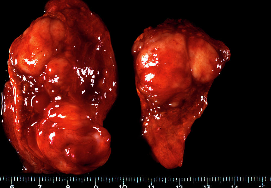
Chromaffin cells: their function is the synthesis and secretion of catecholamines.As it develops from the ectodermal neural crest, it contains two populations of parenchymal cells: It forms the central part of the suprarenal gland. The parenchyma is divided into 2 histologically and functionally different regions the cortex and the medulla.A network of reticular fibers supports the secretory cells and the blood vessels of the cortex and medulla. The stroma: the gland is covered by a connective tissue capsule that sends thin septa inside the gland.

Histological structure of Suprarenal gland it has the same origin as the sympathetic nervous system hence médulla is considered as a modified sympathetic ganglion, where the neurons have acquired an endocrine activity and secrete catecholamines. The medulla: the reddish-brown central layer, which lies deep to the cortex.The cortex constitutes nearly 80-90% of the gland.

It originates from the mesoderm, the same origin as the endocrine cells of the gonads secreting steroid hormones.


 0 kommentar(er)
